The neuroscience research community at Washington University, spanning multiple schools across two campuses, is large, diverse and collegial. Importantly, the scope of work performed by our Training Faculty matches the breadth of modern neuroscientific studies. It spans fundamental mechanisms of brain function and development, to neurological diseases and clinical interventions.

A selection of topics Neuroprep faculty mentors study:
- Pain
- Sleep
- Addiction
- Stroke
- Neuroimmunology
- Behavior
- Brain circuitry
- Parkinson’s disease
- Development
- Nerve repair
- Alzheimer’s disease
- Vision
- Language
- Movement
- Psychological disorders
- Neurogenetics
Faculty investigate these subjects using a range of approaches, including computational biology, device engineering, model organisms, neuroimaging, and clinical trials. They examine questions at all scales, from the microscopic to the whole organism.
Each faculty member maintains active, well-funded research programs and engages in meaningful training and education of students. Neuroprep scholars will briefly rotate through three labs at the beginning of their program and select the best fit for continued research.
Neuroprep faculty mentors are affiliated with 15 Departments:
| Anesthesiology | Biology | Biomedical Engineering |
| Cell Biology & Physiology | Developmental Biology | Electrical & Systems Engineering |
| Genetics | Neurology | Neuroscience |
| Neurosurgery | Ophthalmology | Pathology & Immunology |
| Psychiatry | Psychology & Brain Sciences | Radiology |
While doing research, Neuroprep participants will benefit from the synergistic activities of 35 Research Centers, more than 50 Core Facilities that provide services and expertise including:
- imaging of single molecules, cells, circuits and brains
- behavioral phenotyping in small animals and in humans
- genome editing
- biomedical informatics
- tissue culture and stem cell production
- proteomics
- mass spectrometry
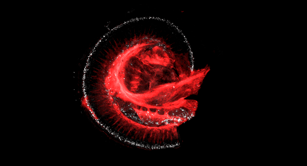
The images on this page were part of the 2021 Sci-Art Competition held by the Department of Neuroscience at the School of Medicine.
Starry Hippocampus, by Yifan Wu, graduate student, Papouin Lab [header image at the top of page]. This is a confocal tile image of mouse hippocampus. There are four different labels: blue labels the excitatory neurons, cyan labels the neuronal nuclei, red labels the astrocytes and green labels all the cell nuclei.
Nautilus Universe, by Rui Feng, PhD, postdoctoral researcher, Cavalli Lab. Immunofluorescence of whole mount adult mouse cochlea was performed to stain for neurons (TUJ1 in red) and satellite glial cells (FABP7 in grey).